Foundations
Gymnastics Anatomy
The Hip & Pelvis: Where the Trunk Meets the Legs
Understanding how a central body region helps organise shape, balance, force and movement
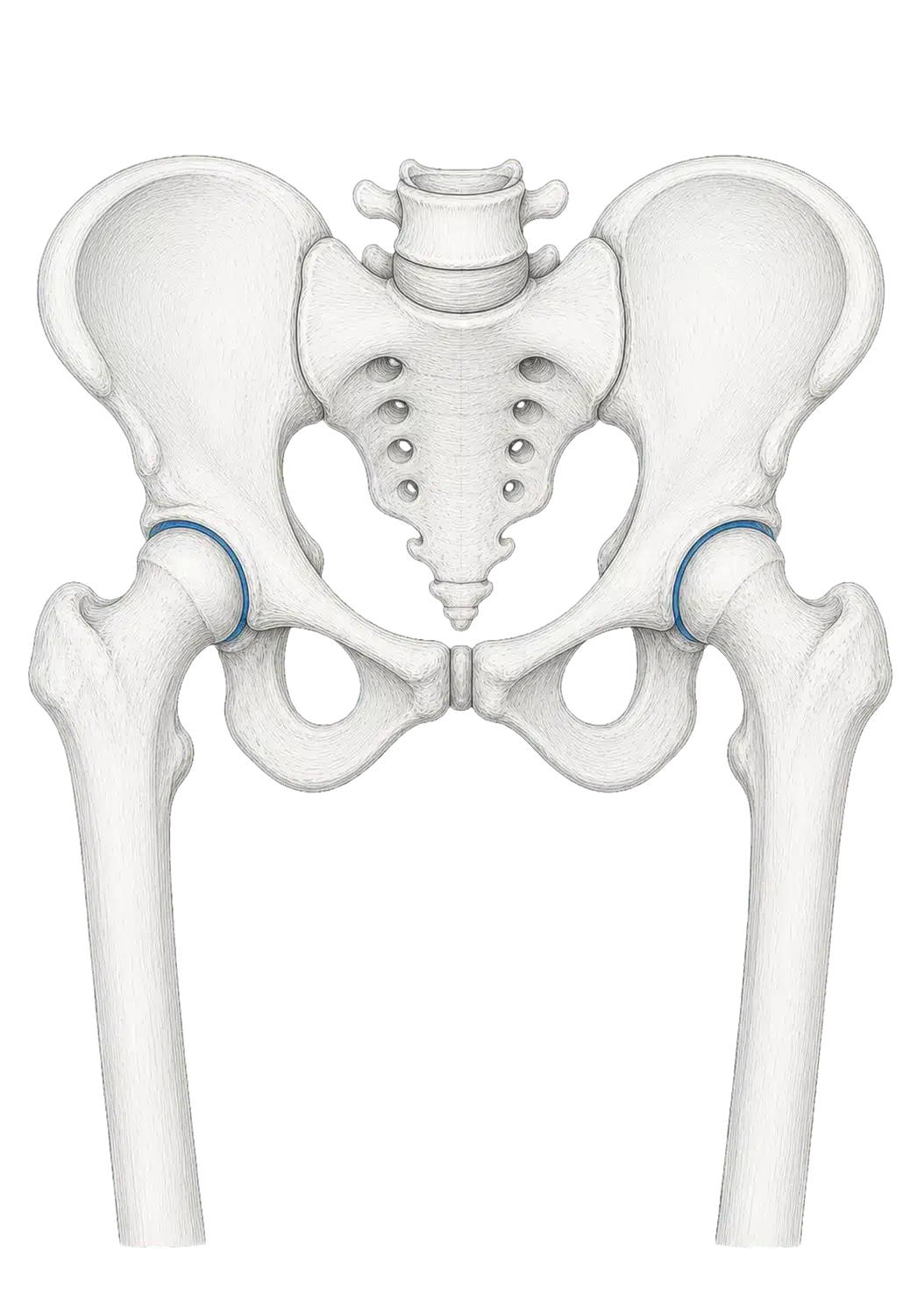
When families hear the word hip, they may think of a single joint at the side of the body. In movement, however, the picture is broader. The two hip joints sit within the pelvis, surrounded by muscles that connect the trunk and legs. Together, this region helps a gymnast change shape, balance on one leg, lift or lower the legs, jump, land and move between positions.
The pelvis is not simply a fixed bowl, and the hips do not work as two isolated hinges. Position, speed, direction and support all change what the region is being asked to do. A quiet balance has different demands from a fast jump. A pike has different demands from an arch. Even when two gymnasts appear to make the same shape, their bodies may organise it slightly differently.
This chapter introduces four important parts of the region: the gluteus maximus; the gluteus medius and minimus; the iliacus and psoas major; and the adductor group. These structures matter, but none works alone. Muscles share tasks with one another and respond to the rest of the body.
The regional concept
A Region That Changes With the Task
The hip and pelvis are involved whenever the trunk and legs have to organise movement together. That contribution changes from task to task.
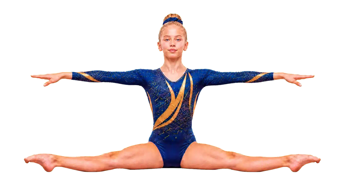
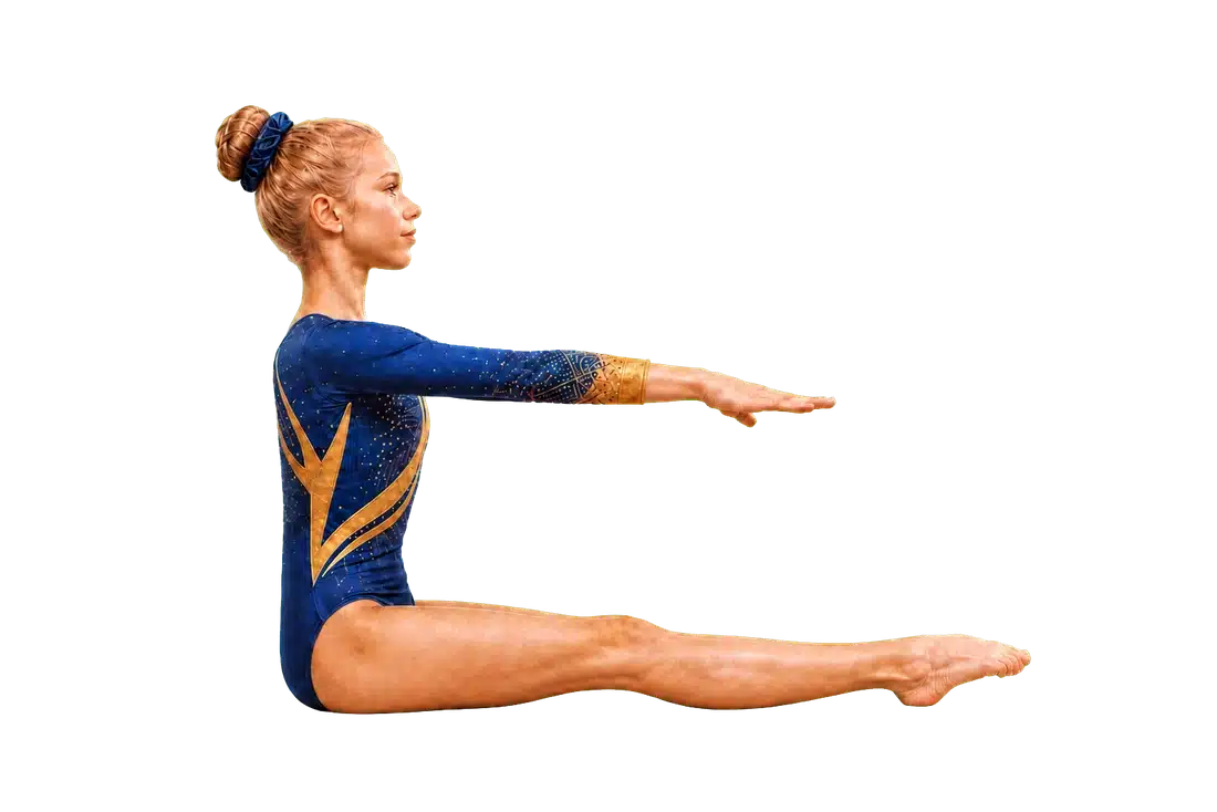
In a two-footed landing, the body is managing motion and force across several joints. The hips, knees and ankles change position while muscles around the trunk and lower limbs contribute to control. In a single-leg balance, the demand is different: the body must organise itself over a narrower base of support. In a pike or straddle, range of motion becomes more visible, but range is only part of the story. Control, timing and the position of neighbouring joints also matter.
This is why anatomy cannot be reduced to a list in which one muscle has one job. A muscle may contribute differently as joint position changes. Several muscles can share a task. The same muscle can shorten, lengthen or hold relatively steady while producing or controlling force.
Position is not performance
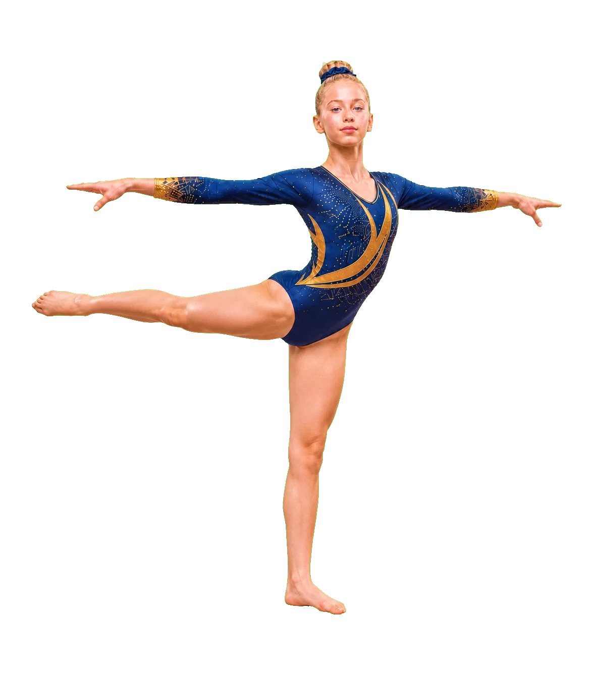
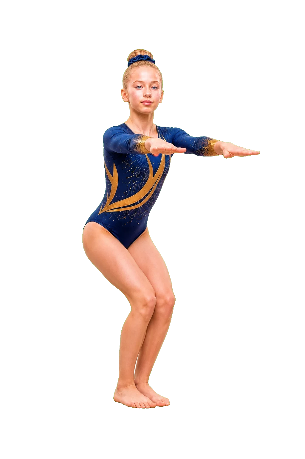
A still photograph captures only one instant. It cannot show how a gymnast arrived there, what forces were present or how the position was controlled. Two shapes that look similar may have been produced through different combinations of movement at the hips, pelvis and spine.
Stability is not stiffness
In family conversations, stable can sound like motionless. In movement science, stability is better understood as the ability to organise and adapt. Sometimes the pelvis moves. Sometimes it remains relatively steady. What matters is whether the movement suits the task—not whether the body has been frozen into one universal posture.
Foundational anatomy
What Do We Mean by the Hip and Pelvis?
The pelvis is a ring of bones at the base of the trunk. On each side, the ilium, ischium and pubis form part of a socket called the acetabulum. The rounded head of the femur—the thigh bone—fits into this socket to form the hip joint[1].
Because the hip is a ball-and-socket joint, it can move in several directions. The thigh can move forwards and backwards, away from and towards the midline, and rotate. Most real movements combine these possibilities rather than staying perfectly within one plane.
The pelvis also meets the spine at the sacrum and connects the left and right sides of the body. It therefore belongs to both the trunk and lower-limb story. Muscles cross from the pelvis to the femur, from the spine to the pelvis or femur, and across neighbouring regions. Their pull depends partly on where the bones are positioned.
A joint built for movement and support
The shape of the femoral head and acetabulum gives the hip substantial bony stability while still allowing a wide range of motion[1]. The joint capsule, ligaments, labrum and surrounding muscles also contribute. This chapter concentrates on muscles, but muscle is only one part of the system.
The pelvis is not one joint
It is common to talk about “the pelvis” as if it were a single movable piece. In practice, the region includes several bones and joints, and its visible position reflects relationships among the hips, spine and whole body. A change that appears to come from the pelvis may involve more than one place.
Muscle Spotlight
Gluteus Maximus
Where it sits
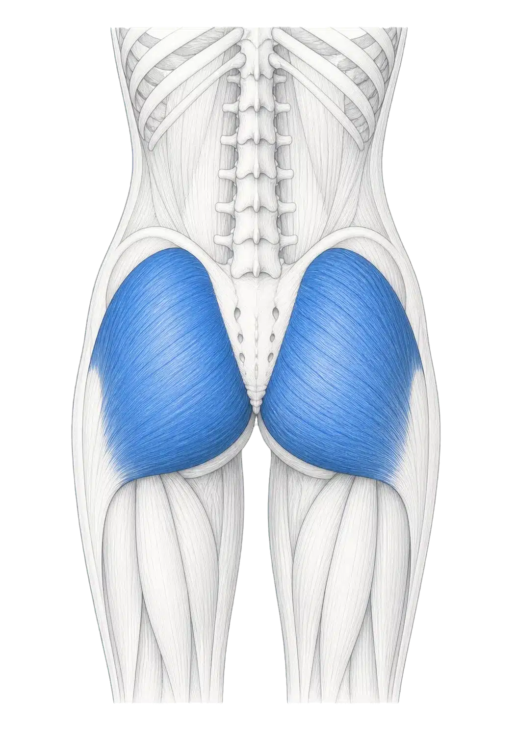
The gluteus maximus is the large, superficial muscle at the back of the hip. It arises across the back of the pelvis and sacral region and attaches to the upper femur and the iliotibial tract.
What it does
It contributes strongly to hip extension—moving the thigh behind the body or helping the body rise when the hip is flexed. It can also contribute to rotation and, depending on position and which fibres are involved, to movements towards or away from the midline.
In gymnastics
Hip extension appears in many forms: driving the body upwards from a more flexed position, moving towards a straight line, controlling a change between shapes, or helping organise the lower body during jumping and landing. The gluteus maximus can contribute, but it does so alongside the hamstrings, adductors, trunk muscles and other structures.
Research examining exercise tasks confirms that gluteus maximus force changes considerably with the movement and external demand[5]. That is useful evidence about task dependence; it is not a ranking of gymnastics exercises for children.
From Anatomy to Movement
Extension is shared work
The gluteus maximus is strongly associated with hip extension, but the body does not allocate the entire action to one muscle. The hamstrings cross the back of the hip, and part of the adductor magnus can also contribute to extension. Their relative contribution changes with joint position, speed, load and whether the feet or hands are supporting the body.
In gymnastics
- moving from a crouched position towards standing;
- preparing and producing force for a jump;
- changing from a more folded shape towards a straighter line;
- controlling the hip as the body receives force in a landing;
- contributing to an arch or leg-behind-body position.
These examples share anatomy, but they are not the same task. A landing involves absorbing and controlling motion. A jump involves producing motion. A held shape may involve relatively little visible movement even while muscles produce force.
The body does not read muscle diagrams
Anatomy illustrations separate structures so that we can see them. The nervous system does not activate a page in a textbook. It coordinates many muscles across many joints, continuously adjusting to balance, direction, speed and the environment.
Muscle Spotlight
Gluteus Medius & Minimus
Where they sit
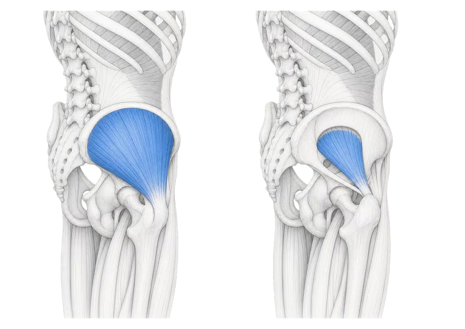
The gluteus medius lies on the outer surface of the pelvis, partly covered by the gluteus maximus. The smaller gluteus minimus lies deeper. Both attach from the pelvis to the upper femur.
What they do
They contribute to moving the thigh away from the body's midline—hip abduction—and to controlling the relationship between the pelvis and femur. Different portions have different lines of pull, so describing either muscle as having only one action is too simple.
In gymnastics
The lateral gluteals may contribute when a gymnast:
- balances on one leg;
- moves the legs apart in a straddle position;
- controls side-to-side organisation;
- changes direction;
- manages the relationship between the pelvis, thigh and knee during take-off or landing.
The contribution is shared with other muscles. A visible shift of the pelvis is not, by itself, proof that these muscles are weak.
Research on gluteus medius shows that its segments can behave differently across exercise conditions[4]. This supports a broader lesson: muscles are not uniform switches, and task context matters.
Movement interpretation
Stable Does Not Mean Still
Pelvic stability is adaptable control.
The word stability often brings to mind a body held rigidly in place. Human movement works differently. The pelvis may remain relatively steady in one task and move substantially in another. Stability describes the ability to manage position and movement in relation to the demand.
In a balance
Small adjustments are normal. The foot, ankle, knee, hip, pelvis, trunk, arms, head and vision all contribute. A gymnast may appear almost still while the body makes continuous corrections.
In a jump or landing
The pelvis changes position as the hips and knees flex and extend. Preventing all movement would not be the goal. The body needs an organised way to produce or receive force.
In a shape
The visible line reflects movement across more than one joint. A pike, arch or straddle cannot be explained by pelvic position alone.
Control is not a personality trait
Children do not either “have” or “lack” control. Coordination develops through experience, instruction, growth and practice. It can look different across skills and from one day to another. Fatigue, attention, confidence and unfamiliarity can all influence what families see.
Muscle Spotlight
Iliacus & Psoas Major
Where they sit
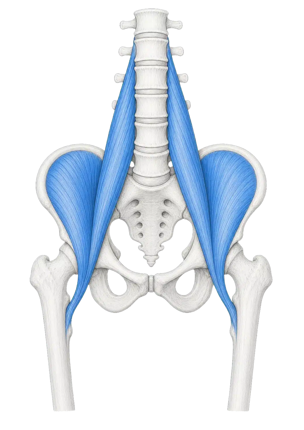
The iliacus begins on the inside of the pelvis. The psoas major begins along the lumbar spine. They travel through the front of the hip and attach near the upper femur. Together they are often discussed as the iliopsoas, although modern anatomical work shows that their structures and insertions should not be treated as perfectly identical[2].
What they do
Both contribute strongly to hip flexion—bringing the thigh and trunk closer together at the hip. Their position also allows them to contribute to hip and trunk organisation in ways that depend on the task and which part of the body is moving.
In gymnastics
Hip flexion is visible when a gymnast:
- lifts a leg forwards;
- moves towards a pike;
- brings the thighs towards the trunk;
- holds or changes a compressed shape;
- steps or runs before a gymnastics action.
These muscles work with other hip flexors, including rectus femoris and sartorius, and with the abdominal and thigh muscles. The visible movement is never the work of iliopsoas alone.
A common misunderstanding
Flexion Does Not Automatically Mean “Tight”
Gymnastics uses large ranges of motion, so families often hear words such as tight, flexible and open. These words can be useful in ordinary conversation, but they are not precise diagnoses.
A feeling is real—but the explanation may be uncertain
A child may describe a pulling, effort or restriction around the front of the hip. That experience matters. It does not, on its own, identify which structure is involved or why the sensation occurs. Joint position, unfamiliar movement, muscle effort, fatigue, growth, previous activity and many other factors can influence how a movement feels.
Range is not controlled by one muscle
Hip movement reflects the shape of the bones, the joint and surrounding tissues, the position of the pelvis and spine, nervous-system tolerance, familiarity with the task and active control. Stretching one named muscle is not a universal solution.
More range is not always the goal
Gymnastics sometimes rewards large ranges, but useful movement also requires strength, timing and control within the range a gymnast uses. A child should not be pushed towards a target because another gymnast appears more flexible.
Muscle Spotlight
The Adductors
Where they sit
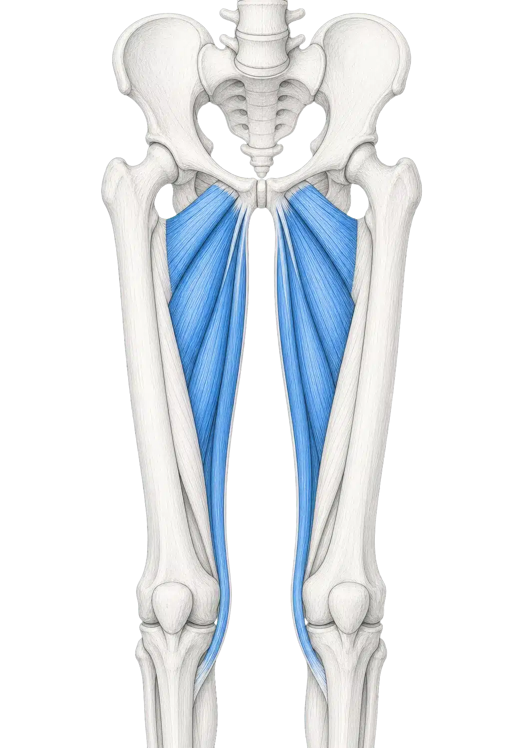
The adductors are a group of muscles along the inside of the thigh. The group includes adductor longus, brevis and magnus, together with gracilis and pectineus in common functional descriptions. Their origins, insertions and lines of pull differ.
What they do
Their name comes from adduction—moving the thigh towards the body's midline. That is only part of the story. Depending on the muscle and joint position, members of the group may also contribute to hip flexion, extension or rotation. In the transverse plane, a published gait model describes roles in producing or controlling femoral rotation at different phases[8].
In gymnastics
The adductors may contribute when a gymnast:
- brings the legs towards one another;
- controls movement into or out of a straddle;
- organises the thighs in a straight-body position;
- transfers force between the pelvis and legs;
- stabilises or changes direction during support, take-off or landing.
From anatomy to movement
More Than Squeezing the Legs Together
The adductors are a useful example of why muscle names can mislead. Their group name describes one direction of movement, yet their actual contribution depends on geometry and task.
In a straddle
As the legs move apart, tissues on the inner thigh may lengthen while also producing force to control the movement. When the legs return towards the midline, the same region may contribute differently. The body is not simply “on” in one direction and “off” in the other.
In a jump or landing
The thighs, pelvis and trunk must organise force in three dimensions. Hip adduction is only one visible component. Rotation, flexion and extension, together with knee and foot position, change the demand.
In a straight shape
When coaches ask for the legs to remain together, the cue concerns the whole visible line and the organisation of the movement. It does not mean that the adductors should be contracted maximally at every moment.
Why joint position matters
As the hip bends, straightens or rotates, a muscle's line of pull relative to the joint changes. Biomechanical models therefore sometimes disagree with the one-word actions printed in basic anatomy lists. This is not a failure of anatomy. It is a reminder that living movement is more complex than a table.
Movement Lens
Straddle, Pike, Balance & Landing
These familiar situations show how the hip and pelvis change their organisation with the task.
Straddle
The thighs move away from the midline, with position shared among the hip joints, pelvis and trunk. The lateral hip muscles and adductors can both contribute, but in different ways and at different moments. A wider straddle is not automatically a better or safer one.
Pike
The hips flex as the thighs and trunk move closer. The hip flexors may help create or hold the position, while other muscles control the trunk, knees and ankles. The visible shape does not tell us how much movement occurred at each joint.
Single-leg balance
The body organises itself over one hip and one foot. Gluteus medius and minimus may contribute to the relationship between pelvis and femur, while the trunk, standing leg, foot, arms, head and vision all participate.
Landing
The hips, knees and ankles change position as the body receives force. Muscles around the hip contribute to controlling motion, but landing is a coordinated whole-body action. Gymnastics research shows that landing patterns vary with age and training stage, and findings from specific laboratory tasks should not be turned into one universal technique rule[7].
The hip and pelvis do not need to look the same in every task. Good organisation means responding appropriately to the movement, the surface and the gymnast.
What Changes Between Positions?
The direction of movement
A straddle emphasises movement away from the midline. A pike emphasises hip flexion. A balance may involve very little visible motion. A landing includes rapid changes across several joints.
The base of support
Two feet, one foot, the hands or an apparatus create different relationships between the body and the environment. The hip muscles respond within that larger support system.
The amount of force
A gentle shape on the floor differs from a dynamic take-off or landing. A position that appears similar can place very different demands on the body depending on speed and external force.
The role of gravity
The same joint action can require different muscular contributions when the body changes orientation. Lifting a leg, lowering it or holding it against gravity are related but distinct tasks.
The available range
Children vary. Hip structure, age, experience, confidence and other factors influence the range they use. Research in typically developing children demonstrates variation in measured hip range and cautions against treating one value as a universal norm[6,10].
The need for precision
Some tasks allow more individual variation; others require a narrower relationship with an apparatus, partner or landing area. Coaches adapt cues to the purpose of the movement.
The family view
What Families May Notice Over Time
Children's movement changes as they grow, learn and become familiar with gymnastics. Families may notice:
Some positions become easier to organise
A child may begin to understand where the hips are and how a cue relates to a shape. This can look like a change in strength or flexibility even when learning and confidence are important parts of the improvement.
Range does not develop evenly
One direction may feel easier than another. Left and right sides may not look identical. Small differences are common and do not, by themselves, establish a problem.
Growth can change familiar movement
Changes in limb length, body proportions and body mass can temporarily alter timing and the way a familiar skill feels. The child is not necessarily “going backwards”; the movement system may be adapting.
The same child can look different on different days
Sleep, school demands, travel, food, mood, attention, recent activity and confidence can all influence readiness. A single lesson is not a complete measure of development.
Coaching language becomes more meaningful
Words such as hips, square, straight, pike and straddle become connected to repeated movement experiences. Understanding develops gradually.
The evidence boundary
What the Science Allows Us to Say
Established anatomy is the strongest foundation
The femoral head and acetabulum form the hip joint, and muscles around the pelvis and femur contribute to movement and control[1]. The broad locations and actions described in this chapter are well established.
Muscle function is task-dependent
Modern anatomical, electromyographic and modelling research shows why a muscle cannot always be assigned one fixed action. Joint position, force direction, speed and the rest of the body change its contribution[2,4,5,8].
Gymnastics research adds context—but has limits
Laboratory studies can compare landing forces, joint angles or muscle activity. One study involving gymnasts and age-matched non-athletes found different neuromuscular landing patterns across training stages[7]. Its specific task, sample and measurements matter. It does not tell us how every child should land, nor does it isolate the hip and pelvis from the rest of the body.
Children's bodies show variation
Paediatric studies demonstrate variation in hip range of motion and developmental measures[6,10]. These data help clinicians and researchers describe groups. They should not be converted into magazine flexibility targets or used to judge a child from a photograph.
What the evidence does not support
The research reviewed here does not support diagnosing a child as having “inactive glutes”, a “tight psoas”, a “misaligned pelvis” or a specific injury based on an ordinary family observation. It also does not establish one ideal pelvic position for every gymnastics task.
Anatomy helps us ask better questions. It does not remove the need to observe the whole child and the whole task.
Safety and Health Notices
This chapter is educational. It is not an assessment, diagnosis, treatment plan or individual flexibility programme.
Gymnastics positions can create strong sensations of effort or stretch. Children should be encouraged to describe what they feel in clear, ordinary language. Coaches and families should not pressure a child through pain or use the magazine to decide which structure is responsible.
Stop and communicate
A child should tell the coach if a movement causes pain, feels suddenly different or makes it difficult to continue normally. The coach can stop or adapt the activity and inform the parent or carer in line with the programme's procedures.
Seek appropriate assessment
NHS guidance says hip pain in children should be checked because several causes are possible. Sudden hip, thigh or knee pain, limping or inability to put weight on one leg requires urgent advice through a GP or NHS 111[9]. Always follow current NHS guidance and local medical advice.
Avoid home diagnosis
Posture, side-to-side difference, a limited-looking shape or a feeling described as “tight” cannot identify an injury. Do not use stretching, massage or strengthening to treat an assumed problem without appropriate guidance.
Return belongs to the individual
After pain or injury, return to gymnastics should follow advice from the appropriate healthcare professional and communication with the coach. A magazine cannot set a return timetable.
References
- Glenister, R., & Sharma, S. (2023). Anatomy, bony pelvis and lower limb, hip. In StatPearls [Internet]. StatPearls Publishing. Official NCBI record. PMID: 30252275.
- Sedlmayr, J. C., Bates, K. T., Wisco, J. J., & Schachner, E. R. (2022). Revision of hip flexor anatomy and function in modern humans, and implications for the evolution of hominin bipedalism. The Anatomical Record, 305(5), 1147–1167. doi:10.1002/ar.24769. PMID: 34569157.
- Tramer, J. S., Hölmich, P., & Safran, M. R. (2024). The iliopsoas: Anatomy, clinical evaluation, and its role in hip pain in the athlete: A scoping review. Journal of the American Academy of Orthopaedic Surgeons, 32(13), e620–e630. doi:10.5435/JAAOS-D-23-01166. PMID: 38502896.
- Ebert, J. R., Edwards, P. K., Fick, D. P., & Janes, G. C. (2017). A systematic review of rehabilitation exercises to progressively load the gluteus medius. Journal of Sport Rehabilitation, 26(5), 418–436. doi:10.1123/jsr.2016-0088. PMID: 27632888.
- Collings, T. J., Bourne, M. N., Barrett, R. S., Meinders, E., Gonçalves, B. A. M., Shield, A. J., & Diamond, L. E. (2023). Gluteal muscle forces during hip-focused injury prevention and rehabilitation exercises. Medicine & Science in Sports & Exercise, 55(4), 650–660. doi:10.1249/MSS.0000000000003091. PMID: 36918403.
- Sankar, W. N., Laird, C. T., & Baldwin, K. D. (2012). Hip range of motion in children: What is the norm? Journal of Pediatric Orthopaedics, 32(4), 399–405. doi:10.1097/BPO.0b013e3182519683. PMID: 22584842.
- Niespodziński, B., Grad, R., Kochanowicz, A., Mieszkowski, J., Marina, M., Zasada, M., & Kochanowicz, K. (2021). The neuromuscular characteristics of gymnasts' jumps and landings at particular stages of sports training. Journal of Human Kinetics, 78, 15–28. doi:10.2478/hukin-2021-0027. PMID: 34025860.
- Leighton, R. D. (2006). A functional model to describe the action of the adductor muscles at the hip in the transverse plane. Physiotherapy Theory and Practice, 22(5), 251–262. doi:10.1080/09593980600927385. PMID: 17118893.
- NHS. (2024, April 3). Hip pain in children (irritable hip). Official NHS guidance.
- Wong, C., Petersen, M. M., Henriksen, T., Jurca, A., Boedtker, S., Balslev-Clausen, A., & Harsted, S. (2025). Angular alignment, rotational profile, and joint range of motion in the lower limb of typically developing children from 7–16 years of age: A cross-sectional study. Acta Orthopaedica, 96, 363–370. doi:10.2340/17453674.2025.43478. PMID: 40322978.